What is the labrum?
The labrum is a type of cartilage that attaches to the socket of the ‘ball and socket’ hip joint. This provides extra stability to the hip. A Labral Tear can be as the result of an accident/injury or due to degeneration, or repetitive friction (hip impingement).

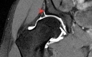
Picture A – MRI arthrogram of the hip – the arrow is showing Contrast behind the labrum; this can only happen if the labrum is torn
How does it tear?
The Labrum can tear either as a result of a
- Chronic injury from repetitive use and activity. These tears are referred to as degenerative tears or a labral detachment. This is the most common mechanism, and results from hip impingement where excess bone on the ball of the hip (a spur or CAM lesion) rubs against the labrum and causes a tear or detachment. At this point you will have symptoms.
- Acute injury following a fall or sports injury. These are referred to as traumatic tears and are commonly the result of sudden twisting movements.

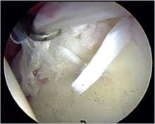
Picture B – Arthroscopy picture showing torn Labrum
Who is at risk of a Labral tear?
Labral tears are more common in women. People with pre-existing hip abnormalities, like dysplasia or extra bone around the hip joint are also at greater risk. People whose sport requires regular rotating of the hip (golf, ballet and football) also have an increased risk.
What are the symptoms of a Labral tear?
The most common symptoms patients report are;
- Hip pain
- Clicking or catching
- Stiffness
- Locking
You will have groin pain and this will be worse in any movement where the hip is flexed and rotated, e.g. performing a squat, getting in and out of a car, or repetitive movements e.g. running.
How would it be diagnosed?
During your initial appointment, the consultant will take a history from you. This will cover any injuries you may have sustained to the hip, any previous surgery you may have had and a record of your symptoms. This history along with a physical examination will help your consultant with his diagnosis and help them determine which imaging would be best to confirm the suspected diagnosis.
If a labral tear is suspected, your consultant is likely to request an MRI arthrogram – Picture A is an example of what the consultant will be able to see from an MRI arthrogram. These images help the Surgeon identify if there is a Labral tear and also the degree of the tear. These images can also be used to assess the rest of the hip, and exclude anything else that may be going on in and around the joint.
How is a Labral Tear treated?
Conservative treatment with tablet analgesia is the first line, but many patients would have tried this before consulting an orthopaedic surgeon.
Physiotherapy is used to maximise hip strength and stability, whilst teaching you to avoid positions and movements that would worsen your symptoms. Activity modification is important: reducing certain activities (e.g. running), avoidance of deep squats.
Hip injections (under local or general anaesthetic) can be used for diagnostic and therapeutic reasons; this is performed in the operating theatre using X-ray control.
If symptoms persist or are severe then surgery is indicated. Key-hole surgery (arthroscopic) is performed on the hip joint to assess and repair the Labral tear. The tears can be stitched back together or removed as necessary. Sometimes there are loose bodies (from the surrounding bone and tissues) in the hip joint that are causing pain and reducing movement – these can be removed at the same time. At the same time, the cause for the tear is assessed and removed (most commonly excess bone formation around the hip joint).
When would I get back to normal?

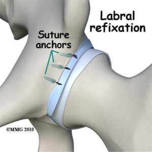
Image showing how the labrum is repaired using suture anchors that fix into the bone

After surgical reconstruction of a Labral Tear, most people return to their normal daily activities in 4-6 weeks. They usually return to sport within three to six months after the surgery. The rehabilitation process is key to a successful outcome following surgery.